
| Technology For Health - 2 |
March 24, 2000 - BBC
A revolutionary surgeon's scalpel can analyse the tissue it is cutting and tell whether it is cancerous or normal.
This could help doctors minimise the amount of healthy tissue removed during operations - and make sure tumours have been completely cut out.
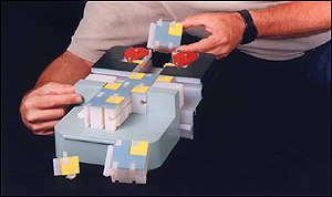
The "smart scalpel", developed by scientists at the US Department of Energy's Sandia National Laboratories, uses "nanotechnology" to check individual cells for signs of cancer.
The cells are pumped from near the cutting edge through a tiny laser.
Cancerous cells have a different chemical makeup to normal cells and change the laser light which passes through them.
This allows a computer to distinguish between the two.
Untested in real operations
So far the technology has only been tested successfully on cancer cells grown in the controlled conditions of the laboratory.
In a real operation, it would have to cope with a large amount of unwanted debris, along with cancer cells which appear very different to those grown in a petri dish.
Professor Garth Cruickshank, a professor of neurosurgery at Queen Elizabeth Hospital in Birmingham, said the idea of a "smart scalpel" was interesting, although he said he could not predict how much difference it would make to the survival of patients.
"Unless you get every single cancer cell removed - which you never do - other factors, such as the responsiveness of the tumour to chemotherapy or radiotherapy, will always be more important."
The cells involved were human brain cells and their malignant equivalents, called glioblastomas.
But those developing it believe it could help neurosurgeons accurately remove tumour tissue obscured by blood, muscle and fat.
Paul Gourley, who leads the project, said: "We can quickly identify a cell population that has abnormal protein content, as do tumour cells, by passing only a few hundred cells - a billionth of a litre - through our device.
February 19, 2000 - UPI - Washington
Scientists hope to use satellite data for rainfall and vegetation patterns to predict conditions for disease outbreak, according to a group of scientists at Johns Hopkins School of Public Health in Baltimore, Md.
Jonathan Patz, a scientist at the school, said satellite data can be used to predict outbreaks of hantavirus, which is a deadly rodent-borne disease, and diarrhea. In years with unusually heavy rainfall, mice thrive due to the lush vegetation. Using satellite data, Patz and his team were able to locate areas where residents were unaware of the threat of the rodent-borne disease. Patz was speaking Friday at the annual meeting of the American Association for the Advancement of Science.
Patz is also tracking satellite images of the Chesapeake Bay to see if he can identify conditions that trigger the growth of cryptosporidia, a water-borne bacteria that causes severe diarrhea and can be deadly to the young, elderly and those with compromised immune systems. Most water treatment plants cannot eliminate the bacteria from the water they process.
"We are coming closer to an ultimate goal of predicting, and ultimately preventing, disease outbreaks," said Rita R. Colwell of the National Science Foundation in Arlington, Va.
Colwell and her colleagues have linked cholera epidemics to environmental changes that can be picked up on satellite images. When sea surface temperatures rise, tiny plants and animals called phytoplankton and zooplankton grow faster. Vibrio cholerae, the bacterium that causes cholera, lives among the plankton. As the temperature of the water rises, so does the sea level, sweeping the cholera-containing plankton into the estuaries where people collect drinking water. The infected water can trigger disease epidemics.
Satellite data can help researchers monitor ocean temperatures to predict cholera outbreaks, follow heavy rainfall and associated flooding to track diarrhea, or prime conditions for mosquitoes, which cause a variety of diseases, Patz said. Earlier this year, a team of researchers reported that they used satellite data to predict outbreaks of Rift Valley fever, a mosquito-borne disease prevalent in parts of Africa.
"Waterborne infections account for 80 percent of all infectious diseases worldwide," said David Pimentel of Cornell University in Ithaca, N.Y. "Climate change, deforestation, chemical pollution and other causes of environmental degradation also contribute to increasing disease incidence in humans."
Infectious diseases may not be the only health problems linked to climate change, the researchers said. Heat waves can trigger deaths due to heat stress; air pollution can set off asthma and other lung diseases; climate change may affect a country's food and water supplies.
"Climate change may lead to a pattern of intensity and duration of extreme events," said Paul R. Epstein of Harvard Medical School.
Such weather extremes are conducive to disease, he said. For example, flooding fosters fungal growth, cholera, and cryptosporidia.
Global warming affects higher latitudes more than lower latitudes, and is more pronounced at higher elevations than lower ones. These new temperature gradients affect the pressure of the air masses and can set up new patterns that pull in different winds and weather patterns, changes that may then affect the diseases endemic to an area, Epstein said.
November 29, 1999 - AP
FONAR Corporation - The Open MRI Company�, announced plans to introduce The Stand-Up MRI�, which will provide for the first time MRI images of the patient in the weight-bearing state. This technology is a superior diagnostic tool for spines and joints and is the first of its kind in the MRI industry. "It will be especially valuable for a host of different applications in sports medicine," said Dr. Raymond Damadian, president and founder of FONAR.
The unique design of The Stand-Up MRI� enables full range-of-motion movements of any joint in virtually any direction, especially important in the diagnosis of sports injuries, but an impossibility in any other MRI scanner on the market. Cardiovascular MRI evaluations can also be performed when the patient is standing or when the patient's feet are elevated above his or her head in the Trendelenburg posture.
Images from The Stand-Up MRI� will be shown for the first time on this works-in-progress scanner, at the Radiological Society of North America (RSNA). The RSNA meeting is the world's largest medical trade show and starts on November 28 in Chicago.
"Some 50 percent of MRI scans are conducted on the spine," said Dr. Damadian. "This is a truly a remarkable diagnostic tool and the need for this one-of-a-kind product is great."
"The Stand-Up MRI� will be ideal for trauma centers where cardiovascular evaluations are key and where immediate and easy-access MRI screening can improve a patient's chances for survival," said Dr. Damadian.
In 1980, FONAR introduced the world's first commercial MRI whole-body scanner. The widespread application of Magnetic Resonance Imaging (MRI) in medicine and biology today is the direct result of the landmark discoveries and early pioneering work of Dr. Damadian. His seminal discoveries of the variations in soft body tissue relaxation times, as well as the cancer detecting NMR signal, are the means used by all MRI scanners to detect cancer and distinguish healthy versus diseased tissues. A 1997 U.S. Supreme Court ruling affirmed that Dr. Damadian's patented discoveries are fundamental to every MRI unit in the marketplace today.
November 11, 1999 - AP
Researchers at the University of Florida Brain Institute "downloaded" the world's most powerful imaging magnet for research -- to be used in investigating a long list of brain and spine-related diseases and injuries.
Weighing 24 tons and carrying a price tag of $2.4 million, the magnet was hoisted gingerly from a semi truck by a construction crane and lowered through the opened roof of a steel-encased building at the UF Brain Institute.
An additional $700,000 to $800,000 worth of computers and ancillary parts will be installed over the next two months before the system becomes operational.
The new magnet has a unique combination of 11.7-tesla magnetic field strength -- 234,000 times stronger than the Earth's natural magnetic force -- and a 40-centimeter cylindrical chamber that will accommodate anesthetized animals up to the size of 15- to 18-pound primates. The next most powerful magnetic resonance imaging (MRI) system, located at the University of Minnesota, has a 9.4-tesla magnet with a 31-centimeter chamber.
"The level of magnetic force makes a big difference in imaging capability, and thus this technology will revolutionize research into all parts of the central nervous system," said Dr. William Luttge, executive director of the campuswide UF Brain Institute.
"This new magnitude of imaging capability will strengthen studies of brain and spinal cord injuries, stroke, epilepsy, tumors and various neurodegenerative diseases, including Alzheimer's and Parkinson's diseases," Luttge said. "With this technology, scientists also will be able to assess the safety and potential benefits of high-powered MRI scanning for clinical use in detecting disease and guiding surgery, as well as the delivery of therapeutic drugs or radiation."
Once the magnet is electrically charged and connected to computers that will direct its functions, it will generate three-dimensional pictures of tissues in animals and in human/animal tissue samples with much finer resolution than can be achieved with the conventional 1.5-tesla MRI scanners used in human medicine.
The U.S. Departments of Defense and Veterans Affairs supported the UF Brain Institute's purchase of the magnet and its components through a 1996 cooperative award of $13.3 million.
UF scientists hope to expand collaboration with both agencies in studies of brain and spine injuries and diseases that affect military veterans.
Dr. Kenneth Berns, UF's interim vice president for health affairs, said the 11.7-tesla magnet will strengthen the pioneering UF-VA studies under way to assess the safety and effectiveness of embryonic nerve tissue transplants in human patients with complicated wounds resulting from spinal cord injury.
The new magnet will enable the researchers to assess, in animal models, what happens to nerve tissues that are implanted at or near the site of a spinal cord injury.
A large number of scientists associated with the Brain Institute, including faculty in the Center for Structural Biology directed by Thomas Mareci, look forward to using the new scanner to achieve greater clarity in medical imaging.
The technology will enable them to:
* generate highly detailed images of structural, chemical and electrical processes taking place inside the brain and spinal cord region in living animals -- without having to sacrifice the animals in order to analyze their organs and tissues;
* see how brain tissue in anesthetized animals responds to various stimuli, including sounds, movement, touch, visual images and the delivery of drugs or other therapies;
* locate and plot the boundaries of tumors and other lesions so therapeutic radiation or surgery can be targeted directly to the site of the problem; and
* measure changes in physiological functions such as blood flow and cell water motion caused by injuries and stroke.
Steve Blackband, a physicist who directs the Brain Institute's Advanced Magnetic Resonance Imaging and Spectroscopy Facility, says the magnet will greatly aid his studies of stroke in animal models. He plans to use it in efforts to define the mechanism and scope of brain and nervous system damage caused by stroke -- information that cannot be obtained with standard clinical MRI scanners.
Through functional imaging in living animals, he hopes to answer questions about how stroke damages brain tissues over time and what happens in the stroke-damaged region after drugs are given in an effort to stop the brain cell deterioration.
Ben Inglis, a UF neuroscientist, is excited by the ongoing opportunities to improve imaging technology to aid discoveries that can be applied to patient care. He is part of a team that has developed techniques for obtaining more information at the cellular level from the images generated through MRI.
The world's most powerful imaging research tool made a long journey to UF -- from the manufacturing plant of Magnex Scientific near Oxford, England, to a port in Miami where it cleared customs inspection before being trucked to Gainesville.
September 9, 1999 - Nature
It has just 78 atoms, took four years to build and it has a spindle that takes hours to rotate but it could be the forerunner of a revolution.
Attempts by scientists to produce molecule-sized machines have produced a toolbox of parts, gears, rotors, switches, turnstiles but no one has produced a molecular motor, until now.
Two molecular motors are reported in the journal Nature.
One was constructed by Dr T Ross Kelly and colleagues of Boston College in Massachusetts. One of his motivations was to understand the molecular motors that are found in all forms of life.
"There are a lot of biological motors in nature, muscles, sperm etc., but although biologists have studied them for many decades they still do not understand how they work on a molecular level. Now we may have a clue."
The diminutive motor consists of 78 atoms arranged in two molecules, a three sided spindle composed of star-shaped molecules and a base plate molecule on which it rests.
"It is a bit like a ratchet, it can turn one way but not the other," says Dr Kelly.
Fundamental processes
The wheel gets the energy required to turn from a molecule called ATP. This is the energy source of biological cells. ATP is a molecule with a lot of internal energy which it can be persuaded to release.
Scientists say that understanding how the motor works will broaden our understanding of many fundamental biological processes.
"Nature tends to conserve solutions," points out Dr Kelly, "it does not solve ten similar problems in ten different ways. It finds a common solution. So our molecular motor may help us understand natural molecular motors in cells."
At the moment the motor is not much good for anything, except to demonstrate a principle.
"It does not turn very quickly," he adds. "It takes several hours for the three-spindled wheel to make one revolution. Our next step is to speed it up."
The other molecular motor reported by Nature has been constructed by German, Dutch and Japanese scientists.
Using a specially assembled carbon molecule, they have been able to make part of it rotate in one direction, taking only four steps to make a complete revolution.
This motor is not powered by ATP but by light and changes in temperature.
June 15, 1999
A large-scale prototype of a computer that could be smaller than a living cell has been designed by an Israeli scientist.
Some scientists believe that, in the future, small biological computers could roam our bodies monitoring our health and correcting any problems they may find.
The prototype has been developed by Professor Ehud Shapiro of the Computer Science Department at the Weizmann Institute of Science.
It is being presented at the Fifth International Meeting on DNA-Based Computers at the Massachusetts Institute of Technology.
In terms of the logic on which it operates, the prototype will behave in a similar way to molecules inside a living cell, a "biomolecular machine".
A living computer
Each cell of our bodies is a collection of machines made out of biological molecules. These molecules can form pulleys and gears to move other molecules around the cell.
Some molecules have the ability to assemble and take apart other molecules. Others gather small molecules and use a template to construct new molecules. In a sense, each of our cells is a complicated city of biological machines all working together.
It is possible that a future biomolecular version of Professor Shapiro's device could lead to the construction of computers, smaller than a single cell, and with the ability to monitor and modify them.
If scientists were ever able to build such a computer, its medical applications would be far-reaching. It could swim in our bloodstream or be attached to specific organs monitoring and supplementing their performance.
"For example, such a computer could sense anomalous biochemical changes in the tissue and decide, based on its program, what drug to synthesise and release in order to correct the problem," says Professor Shapiro.
A Turing machine
Existing electronic computers are based on the architecture developed by John von Neumann in the US in the 1940s. But the new mechanical computer is based on the Turing machine, conceived in 1936 by the British mathematician Alan Turing.
The Turing machine uses the basic concepts of computing, reading and writing one bit of data and performing an action depending upon a program. But although the Turing machine is a general-purpose, universal, programmable computer and is key to the theoretical foundations of computer science, it has not been used in real applications.
Like a Turing machine, Professor Shapiro's mechanical device has a "rule molecule" designed so that the processing of the molecule modifies another molecule in a predetermined way.
To demonstrate the concept, Professor Shapiro has built a 30-centimetre-high plastic model of his mechanical computer. He hopes that the advent of improved techniques for making and assembling molecules will mean the day when his computer could be made is not far off.
If it were built from biological molecules it would measure about 25 millionths of a millimetre in length, roughly the size of a cell component called a ribosome.
A safer and more informative alternative to X-rays appears ready for use, with teeth its first target.
The system harnesses radiation called terahertz rays, which sit between infrared and microwaves in the electromagnetic spectrum.
These can partly pass through many common materials and can reveal both the structure and composition of the target.
Toshiba Research Europe (TRE) is developing the method at the Cambridge Science Park, UK. Its managing director, Professor Michael Pepper says: "These are very early days, but it is clear that Terahertz Pulse Imaging is going to be very important, particularly in areas where X-rays are insensitive."
Safer, smarter
Terahertz rays are non-ionising and are therefore thought to be safer than X-rays. Also, the power levels needed to produce a sharp image are usually lower than the background terahertz radiation encountered in everyday life.
A key advantage of terahertz analysis is that it can be used in two ways. Firstly, it can reveal three-dimensional shape by measuring how the waves are affected by the structure of the target. This is similar to radar.
Secondly, terahertz analysis can be used to identify the material found in the target. This is done by using a range of terahertz frequencies and measuring which ones the material absorbs. This is called spectroscopic analysis.
The first application is expected to be in dentistry. Dr Don Arnone is TRE's project leader and says that one mode of use reveals the thickness of a tooth's enamel and while the other displays the internal condition of the tooth.
"We can construct a three-dimensional image of the tooth and rotate it on the computer screen so the dentist can examine each tooth from the optimum angle," he says.
After dentistry, other body tissues will be targeted for the system's development but non-medical applications are also possible.
TRE has demonstrated the usefulness of terahertz waves in quality control in food processing, semiconductor and computer chip manufacture and its ability to find objects hidden in containers. The recent developments in terahertz technology are due to new, cheaper ways of generating and detecting the waves. To produce them, a semiconductor crystal is bombarded with ultra-fast pulses of visible laser light.
BBC Online - May 19, 1999
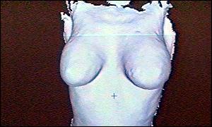
A 3D imaging camera being developed by scientists in Glasgow is set to revolutionise the treatment of cleft lip and breast reconstruction patients. The camera will allow doctors to assess to within a fraction of a centimetre the most appropriate corrective surgery.
Up to now doctors treating babies with cleft palates or women requiring breast reconstruction or reduction have had to rely on experience and guess work during surgery.
The new camera, which mimicks the function of the human eye, can now be used to provide a 3D image which can help them plan reconstructive surgery more accurately and therefore more effectively.
It is particularly helpful in treating wriggly young babies as the image is captured in a tenth of a second.
The camera can also be used to assess facial animation to ensure any operation allows the child's facial expressions to be as normal as possible.
Investigations are continuing into whether the technology can also help detect the first stages of breast cancer.
Although it is still in the research stage, the camera is being used in a number of Scottish hospitals and if a recently submitted lottery application is successful, it could be expanded made available in the rest of the UK.
The director of the Faraday Partnership based at Glasgow University, Dr Paul Siebert, said he believed all hospital equipment will soon be changed to produce 3D images. This would enable doctors to assess the extent of swollen flesh and scar tissue, to name but two examples, and assess the develpment of a disease or even aid diagnosis, he said. He says the digital cameras used for this technique are cheap,fast and effective making them an attractive option.
Dr Ashraf Ayoub-an oral Surgeon at Glasgow's Dental Hospital, says half a dozen children have already taken part in the project although it is too early to tell the results. But he is confident it will help them achieve a near normal look by the time they reach their late teens.
HEALTH INDEX